Introduction
We've covered a lot of ground since Part I, haven't we? And all that's just the tip of the ice berg! Not only is there more information for the advanced student, but it's also a matter of application. It's not enough to know, we also have to do, right? And in that respect, a lot can go haywire. When we're dealing with the creation of fallible human hands, we can divert away from accuracy quite easily if we aren't careful. So translation is another matter entirely.
To that end, let's now discuss details, or those little touches we can add to our sculpture that infuse interest and believability. Details are neither easy to do nor should they be considered afterthoughts. Instead, they should be integrated into our sculpture with equal attention since they can make or break a piece.
We should also understand that many details are fleshy or cornified and so their texture comes into play when we recreate them. We can't just approach them all with the same technique, but need to find those that reproduce how they would feel and look on the real animal. The flesh should appear gooshy and the cornified should appear rough or polished, depending on what they are. For instance, the difference between chestnuts and hoof horn, respectively.
So enough gab...let's go!
Veins
Veins
Veins are patterned and bilaterally symmetrical on either side of the horse, lengthwise. Smaller and finer capillaries obey certain patterns of their own, but are much more random and spontaneous. Both can be very prominent on thin-skinned horses, especially on hot days, or during exercise. Some such horses are the Arabian, Teke, and Thoroughbred.
Since arterial and vein structure is so complex, this section distills the veins into those most obvious on most horses. These primary subcutaneous veins are:
- Facial vein (or submaxillary vein): The “Y” vein that comes from the front tip of the teardrop bone (facial crest) that splits and then branches towards the eye and nostril. It also goes under the jaw, to enter the jugular, and this portion is referred to as the Glosso facial vein; it can produce a subtle visible effect in the throatlatch area.
- Internal subcutaneous vein of the forearm: This major vein of the forelimb is a continuation of the internal metacarpal vein. It passes over the anterior top part of the subcutaneous shaft of the radius and travels to the back of the knee.
- Internal and External saphena vein: A major vein of the hindlimb, it erupts from the groin around the Gracilis and Sartiorus muscles and then branches. The larger anterior branch crosses over the top of the tibia and follows the groove between the front of the tibia and the Tibialis cranialis (Deep flexor metatarsi), over the front of the hock, crossing the top of the metarsal and becoming the internal metacarpal vein. The smaller posterior branch goes towards the Gastrocnemius muscle.
- Internal metacarpal vein: Major vein of the hindlimb and a continuation of the saphena vein.
- Digital veins of the limbs: Pass along the caudal aspects of the limbs into the foot and with two branches, reaches to the front of the limbs. They can be a visible aspect on the horse.
- Spur vein: A major vein of the torso, it often branches into two aspects, an inferior and superior branch. The parent branch lays along the top border of the Posterior deep pectoral muscle and goes into the armpit.
- Subcutaneous abdominal vein: Passes along the posterior of the abdomen and into the posterior of the sternum. It's very distinct on a broodmare, often referred to as the "milk vein" when nursing.
- Capillaries: A series of webbings or "chicken scratches" networking over a region of the body; they are subcutaneous and often very distinct during hot weather or exercise, or on thin-skinned horses. They most commonly manifest on the neck, shoulder, chest, lower haunch, gaskin, and forearm, groin area going up to the anus, with some appearing on front and rear portions of the barrel, or the barrel itself.
Landmarks and Reference Points for Veins
Artistic Aspects to Consider about Veins
Veins should appear squishy and soft, with a rounded surface to blend with the surrounding flesh. They can also have bulbs where they're wider, as often seen with capillaries. The more veins and capillaries we apply, the more we imply heavy exertion or heat dissipation, so we need to make sure our sculpture warrants a heavy or light application of them. Certain breeds with thin skin and "dry" features also tend to exhibit more veins and capillaries such as Tekes, Thoroughbreds, and Arabians. The deft application of veins and capillaries can create a lovely contrast to firm muscle or hard bone, and really drive home the appearance of "living flesh."
Common Artistic Faults
Veins on sculptures are often structured improperly such as being randomly branching when they should be bilaterally symmetrical on either side of the body. This isn't to say they should mirror each other perfectly, but they should present the same general idea in terms of placement and intensity. They also tend to be misplaced, not following their anatomical patterns. For instance, a "Y" vein placed too low on the head or a spur vein placed too high or too low in the elbow area.
They can also be incorrect in structure being indicated as either simply grooves in the clay or flat. They may be clumsily executed and messy, too, due to a shaky hand or poor technique. Size is also a common problem as they can be too big in size or width. The veins and capillaries should also be smooth, and not pebbly, streaky, or rough in texture.
Genitalia
The vulva forms the external opening slit of the urinary and reproductive tracts of the mare. It's approximately four to five inches long and fleshy. The anus is at the top, immediately under the dock, and the vulva is a descending slender oval or hourglass shape. Between the hindlimbs, at the termination of the abdomen, the mare has a fleshy udder comprised of two dangling triangular portions of flesh with a teat on each.
They can also be incorrect in structure being indicated as either simply grooves in the clay or flat. They may be clumsily executed and messy, too, due to a shaky hand or poor technique. Size is also a common problem as they can be too big in size or width. The veins and capillaries should also be smooth, and not pebbly, streaky, or rough in texture.
Genitalia
The vulva forms the external opening slit of the urinary and reproductive tracts of the mare. It's approximately four to five inches long and fleshy. The anus is at the top, immediately under the dock, and the vulva is a descending slender oval or hourglass shape. Between the hindlimbs, at the termination of the abdomen, the mare has a fleshy udder comprised of two dangling triangular portions of flesh with a teat on each.
Between the thighs, at the back of the abdomen, the stallion has a penis, sheath, and two testicles. The penis is enclosed in a double fold of skin called the prepuce that forms the visible sheath outside the body. A normal testicle is egg-shaped or round and about three to five inches long. The size of the testicles vary with individual stallions, but the left is sometimes larger than the right. The testicles are suspended in the scrotum and can be raised or lowered simultaneously or independently.
The vulva, anus, scrotum, and penile sheath are fleshy and relatively hairless.
Landmarks and Reference Points
In both the stallion and mare, the underside of the dock is a good point to gauge where to place the anus. Likewise, where the Gracilis muscles meet along the median seam of the body is a handy reference for placing either the udder or sheath and testicles.
In the mare, the vulva begins about where the points of buttock occur. It can be shaped like a soft oval, more like a rectangle, or have a slight hourglass shape, depending on the natural variation between mares. It should also be straight up and down, and not slanted outwards.
Artistic Aspects to Consider about Genitalia
All genitalia should appear soft and fleshy, not hard. They are passive aspects of flesh (when relaxed) and so are subject to the effects of physics. For example, the udders, sheath, and scrotum jiggle and bounce with motion, or are shifted from side to side. The anus can also pop in and out in synch with the gaits or movements.
Common Artistic Faults
Genitalia is commonly faulted by incorrect size, structure, and placement, even a lack of detail. Many times they're sculpted too harshly, blockish, or blobbish, too, obliterating their soft, fleshy nature. Sculpted vulvas are often slanted, which is a conformation fault. It's not enough to simply pop on blobs of roughly-shaped clay to indicate these features...they need careful shaping and detail just as any other part of the body.
Chestnuts
Each chestnut is distinctive and can sometimes be used to identify animals. They're rough in texture, often having ridges or a pebbly surface. However, they can be softened with oils or lotion, and peeled away to be flatter and smoother, a common practice in show grooming. Sometimes they're oiled for show, which darkens their coloration.
Landmarks and Reference Points
The internal "bump" of the knee is a good point to reference when placing the forearm chestnut whereas the top of the internal splint bone is a good landmark for placing the hindlimb chestnut.
Artistic Aspects to Consider about Chestnuts
Duplicating the cornified texture of a chestnut is important for believability just as much as getting then placed, oriented, and sized correctly. For painting, they can either be a grayish tan, tan, dark brown, dark grey, or nearly black (when oiled), depending on individual variation and the underlying coat color. Often, they're pink or light tan when in a white marking.
While they break off with growth, chestnuts can grow long under natural circumstances to stick out quite a ways from the surface of the arm or hock. This can be commonly seen on wild or feral horses.
Common Artistic Faults
Chestnuts are often misplaced, being placed too high or too low. Or they may be placed bilaterally asymmetrical. They can also be indicated simply by dollops of smooth clay, like smooth buttons, rather than as cornified tissue. They can be too big (often on the hindlimb), too, or not angled correctly (often on the forearm). Sometimes they aren't even present. Mules tend to only have chestnuts on the forearms and not the hind limbs.
Ergots
Landmarks and Reference Points
The flatter aspect of the posterior fore fetlock makes for a useful landmark as does the pointier posterior aspect of the hind fetlock.
Artistic Aspects to Consider about Ergots
Ergots form a gentle, discreet point at the back of the fetlock joint, something especially obvious on a clipped lower leg. They tend to be smoother in texture than the chestnuts. However if allowed to grow, they can become quite pronounced, especially under feathers.
Common Artistic Faults
Ergots are often misplaced or too big.
Whisker Bumps
Whisker bumps or moles occur on the muzzle and around the eye. They vary in size, intensity, and number between individual horses. They can occur a bit randomly or in patterns, again, depending on individual variation and location.
Landmarks and Reference Points
The nostrils and mouth are good landmarks for placing moles on the muzzle. The lower lid is a handy reference for placing those around the eye.
Artistic Aspects to Consider about Moles
Moles are small raised buttons of flesh from which a whisker grows. They're fleshy, squishy, and not hard. They can vary in size on the same horse, or between horses. Those around the eye tend to be smaller than those on the muzzle.
Common Artistic Faults
Moles are most typically flawed by a clumsy rendition that does not make them appear fleshy and soft. They can also be too large and pronounced, lacking a discreet nature. They can also have sunken pits, like a collapsed souffle, or too pointy rather than being smoothly rounded. They can also lack the randomness or pattern indicative of an individual's variation.
Wrinkles
Wrinkles are soft folds of flesh caused by the compression or flexing of skin, or areas of skin that experience a great deal of stretching and movement such as the muzzle. When the animal is standing, wrinkles are most common between the ears, around the eyes, around the muzzle and nostril, in the throatlatch area, at the junction between the neck and the wither, and between the forelegs. In motion, however, wrinkles proliferate as movement dictates.
Wrinkles can vary in size and intensity between individuals. Sometimes certain unusual regions of the body are predisposed to wrinkles, too, such as on the haunch during certain movements, the throatlatch area, or on a heavily crested neck along the crestline or span of the neck. Furthermore, if the animal is laterally bent, large wrinkles on the ribcage may appear on the barrel. Similarly, if the neck is cranked to one side, wrinkles will be apparent on the neck inside the turn, often fanning out onto the shoulder and sometimes the wither area.
Landmarks and Reference Points
Wrinkles occur wherever the flesh is compressed or an articulation has caused the skin to fold. So pay attention to the flesh around articulated joints, or on those areas that are subject to squishing and goo-ing.
Artistic Aspects to Consider about Wrinkles
We should sculpt our wrinkles so they appear as soft, folded flesh. We should also notice that they vary in width and intensity within a "bundle"; they aren't a clone of the one previous. Wrinkles can even wrinkle as clusters wrinkling on each other, such as we often seen in the throatlatch area of a tightly tucked head. Wrinkles also softly fade out into the surrounding skin and don't end abruptly with a definite border.
Wrinkles occur on the muzzle and around the eye, too, with great delicacy and softness. The amount of wrinkling here can vary between individuals, however, with some having few wrinkles while others having a dense proliferation of them.
Wrinkles occur on the muzzle and around the eye, too, with great delicacy and softness. The amount of wrinkling here can vary between individuals, however, with some having few wrinkles while others having a dense proliferation of them.
Common Artistic Faults
Wrinkles are most typically faulted by a clumsy rendition that doesn't make them appear fleshy and soft, but hard and literal. They can also suffer from regimentation, like the folds of an accordion, rather than varying in depth, width, folding, and orientation. They can appear as gouged-out grooves, too, rather than rounded folds of flesh. Wrinkles can end too abruptly as well, rather than softly fading into the surrounding skin.
Secondary Sex Characteristics
Horses have a distinct difference between the genders so a stallion should look like a stallion and a mare look like a mare. Sure there are some exceptions, but on the general scale this works to our advantage in sculpture.
For instance, stallions appear more compact and muscular, often sporting more of a crest and stronger jowls. On the other hands, mares appear "lower to the ground" and longer, with less pronounced jowls. Their necks also tend to be finder and their ears longer.
Geldings, on the other hand, are a bit of a mixture of the two since the full expression of masculinity wasn't allowed to develop. They also often have some eccentricities that disqualified them from being a breeding stallion, and they're fun to inject into clay.
Common Artistic Faults
These characteristics can be overlooked in sculpture so we have a homogenized "horse" rather than a distinct gender. In other words, we can't just swap out "plumbing" and expect to pull off a convincing piece.
Secondary Sex Characteristics
Horses have a distinct difference between the genders so a stallion should look like a stallion and a mare look like a mare. Sure there are some exceptions, but on the general scale this works to our advantage in sculpture.
For instance, stallions appear more compact and muscular, often sporting more of a crest and stronger jowls. On the other hands, mares appear "lower to the ground" and longer, with less pronounced jowls. Their necks also tend to be finder and their ears longer.
Geldings, on the other hand, are a bit of a mixture of the two since the full expression of masculinity wasn't allowed to develop. They also often have some eccentricities that disqualified them from being a breeding stallion, and they're fun to inject into clay.
Common Artistic Faults
These characteristics can be overlooked in sculpture so we have a homogenized "horse" rather than a distinct gender. In other words, we can't just swap out "plumbing" and expect to pull off a convincing piece.
Flesh and Hide, or "Goo"
The term "flesh" entails the muscles and fat beneath the skin. In turn, "hide" refers to the fascia and skin of the animal. Both express themselves, resonating and reacting to motion and articulation.
When it comes to flesh, the novel depressions and concavities of muscles during movement, the amebic absorption of boney parts during certain phases of motion, muscle resonance in response to movement, buckling caused by articulation or force, and the general jiggling and wriggling of flesh are all indicators of gooey flesh that need attention. In terms of hide, the sliding or rippling of skin, its stretched and compressed distortions during movement, and its wrinkles, bumps, stretches and pooches, and soft ridges are also good indicators of "living flesh."
So we can't simply sculpt correct anatomy...we also have to duplicate the nature of flesh as well as express the hide. Being so, it's a common oversight in equine sculpture in lieu of the underlying anatomy. Remember, the hide has been stripped away to reveal the muscular structures in an anatomy chart, but we have to reinstatel it for our sculpture to look real. Truly, a masterly artistic expression of flesh and hide and muscle...goo...is one of the most important ingredients for creating a convincing equine sculpture. What does that mean in practice? It means we should only use our anatomy charts as guides, not gospel. Life presents us with so much more to anatomy than a static chart.


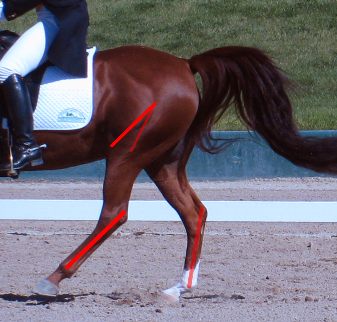
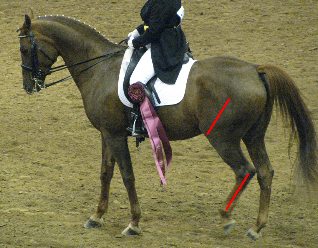
Look how each pectoral is changed away from its resting state simply by walking. Unless we pay attention to muscle morphiing, our sculptures are going to appear artificial and formulaic. Horses don't move like articulated anatomy charts.
Artistic Aspects to Consider about Flesh and Hide
Flesh is soft and inviting, and it depresses and pooches with movement. That means it has a 3D quality, an in and out quality as it goos. Unless we capture this in our sculpture, our muscles will appear flat, contrived, and artificial, more like an articulated flat anatomy chart rather than "living flesh." Similarly, unless we understand and denote the nature of the hide, we're going to create a stylized piece of work that's too polished-looking and hyper-smooth to be truly believable. No horse is smooth like polished metal, but rich in the little imperfections of skin and fascia. Imbuing all this into our sculpture will go far in duplicating a life-like appearance.


Note all the hide and fleshy details to the skin. Horse's aren't "polished" smooth. The smooth finish on Maureen Love pieces is certainly pretty, for example, but it's not realistic. It's a manifestation of artistic style.
Common Artistic Faults
Sculptures are often faulted by a static, formulaic, or stylized rendition of flesh and hide. For example, we cannot apply standing anatomical configurations to a sculpture depicting motion just as much as we cannot apply the anatomy changed by motion onto our standing sculpture. Horses don't move like articulated anatomy charts, with the same configurations occurring over and over again despite anything else, like a "sculpt by numbers" formula. Instead, motion and physics can radically change the nature of the flesh and hide, and we need to capture these changes in our work.
Hooves
Know it or not, hooves are rich in detail from the bars to the clefts to the frog, yet they're often overlooked in sculpture. Detailing the hooves is important to fully express realism. For a more in-depth look at hooves, please refer to the blog post, Steppin' Out: Hooves From An Artistic Perspective.
Common Artistic Faults
Many sculpted hooves are pathological since the artist didn't understand what constitutes a healthy, adaptive, balanced foot. Many sculpted hooves also lack detailing on their palmar side, and usually we're lucky if merely the frog is indicated. But there's so much more to the underside of the foot and we need to factor all those structures into the sculpting of our feet.
Ears and Nostrils
These features are typically hollowed out to enhance realism.
Artistic Aspects to Consider about Ears and Nostrils
Ears are complicated structures, being delicately fluted with a bulbous base. Where the rims meet at the bottom forms a "V" constructed of subtle curvaceous, bulbs, and lip-like structures rather than a literal, simple "V." What's more, these structures change as the ear is rotated, forming rather complex curves, pooches, angles, and overlapping flesh.
The nostril is similarly complicated being so fleshy and gooshy. Being so, the lateral rim stretches and misshapes depending on how its manipulated by the muscles, tendons, and fascia which can distort it well away from its resting aspect. The anterior "comma cartilage" rim can be distorted as well, expanding quite a bit from its resting state. There's also the false nostril that can distort, too, and even form pronounced raised flutes of flesh on either side of the nasal bone. On the side, the flared nostril typically has hollows, curves, and depressions confident to the muscles and tendons that activate them.
Complicating things still further, each nostril can move up or down, or back and forth in relation to its pair, adding to expression, character, and effect.
Complicating things still further, each nostril can move up or down, or back and forth in relation to its pair, adding to expression, character, and effect.
Common Artistic Faults
Errors in the nostril are common. Of particular note is the error of overdoing it with the drill tool. Specifically, the more rounded, bulbous front rim of the nostril may be too thinned as the nostril is hollowed, and sometimes so that front rim can be reduced to a sharp edge. The rims of the nostril can also be sculpted in a clumsy way, creating an uneven, pebbly texture rather than a smooth, fleshy one. On the other hand, the nostrils may be sculpted incorrectly altogether, or with flutes (especially when flared) that don't possess the complex curves and indentions imposed by the overlaying musculature. Nostrils aren't simple things on the horse, and so shouldn't be so straight-forward on a sculpture.
Similarly, equine ears are complicated. Too often, however, they're "scooped out" when hollowed, forming more of a spoon-shape than a fluted one. The bottom "V" is often too simply rendered as well, as though it was simply two rims pinched together (a common sculptural flaw with pre-formed ears). Yet that "V" is characterized by a complicated folding of flesh with bulbs, twists, and overlapping features peculiar to equines alone, and unless our sculpted ears have similarly complex "Vs," those ears don't actually belong to Equus. Just as cat ears and dog ears aren't simple flat triangles (they have those curious folds on their lower, outside rim, where the outer rim meets the head), so a horse's ears aren't simple flutes with a simple "V" on the bottom either.
Mane and Tail
Sculpting the mane and tail (and feathers) is arguably one of the hardest aspects of sculpting realistic equines. It should never be treated as an afterthought. And there are many ways to expressive, but regardless, hair is passive to physics and moves in often complicated, unpredictable, chaotic ways. Its texture also presents a challenge, it being wispy, weighty, striated, and complex.
Mane and Tail
Sculpting the mane and tail (and feathers) is arguably one of the hardest aspects of sculpting realistic equines. It should never be treated as an afterthought. And there are many ways to expressive, but regardless, hair is passive to physics and moves in often complicated, unpredictable, chaotic ways. Its texture also presents a challenge, it being wispy, weighty, striated, and complex.
Artistic Aspects to Consider about the Mane and Tail
The manes and tails on certain breeds can be quite fine and soft, such as on the Arabian. On the other hand, it can be quite coarse such as on the Takhi or Fjord. Similarly, feathers can be silky and wispy on some breeds such as the Clydesdale or Shire, or wiry and dense such as on the Ardennes and Brabant. All this means that different types of hair flow differently, something we should pay attention to. In short, not all hair is created the same.
Hair also has layers; the mane and tail (and feathers) aren't all one equal length. These layers can operate independently or in synch with the other layers, depending on the situation. So unless we think in terms of layers, our manes and tails (and feathers) will appear odd and artificial.
And there are many ways to sculpt hair, given we meet the criteria of its flow and movement, and texture. Some artists merely indicate texture, opting for a more impressionistic approach while others go for a highly detailed rendition, with lots of little striations indicating each hair. It all depends on what we like for our sculptures.
We may also choose to impart waviness such as often seen when manes and tails are taken out of braids that preserve its length. This can be a really appealing touch on a sculpture, especially on Morgans, Andalusians, and Friesians, or other breeds known for a long mane and tail.
Hair also has layers; the mane and tail (and feathers) aren't all one equal length. These layers can operate independently or in synch with the other layers, depending on the situation. So unless we think in terms of layers, our manes and tails (and feathers) will appear odd and artificial.
And there are many ways to sculpt hair, given we meet the criteria of its flow and movement, and texture. Some artists merely indicate texture, opting for a more impressionistic approach while others go for a highly detailed rendition, with lots of little striations indicating each hair. It all depends on what we like for our sculptures.
We may also choose to impart waviness such as often seen when manes and tails are taken out of braids that preserve its length. This can be a really appealing touch on a sculpture, especially on Morgans, Andalusians, and Friesians, or other breeds known for a long mane and tail.
Common Artistic Faults
Missteps here are common. Often we see hair rendered as dreadlocks, tentacles, or "ropes," especially with flowing tendrils. Likewise, we may find the mane sculpted like soft-serve ice cream rather than naturally flowing hair (a flaw I used to do in the past as I was learning). Here's a handy trick: make the tips as thin as possible to make them appear wispy instead of ropey.
Other times, layers haven't been accounted for so we have hair all the same length which gives it an artificial look rather than a natural, flowing one. We also often see texture gouged into the mane and tail (or feathers), often being pill-ed or with torn ridges, as though it was sculpted with a fork. Instead, the hair should be smooth and silky.
Hair is also 3D, so it should have areas of hollows and areas of bunched-up thickness. If we sculpt it all the same, like a 2D rendition, it won't look natural and real.
More still, we find that the sculpted hair lacks the passive, unpredictable movement characteristic of its flow, instead taking on a regimented "safe" expression. Remember, hair is passive to motion, so we need to keep the movements of the body and physics in mind when we design it. And often times, hair can move counter-intuitively to the motion as a result, so pay attention to life study and reference photos.
Even so, we may find that the sculpting of the mane and tail (and feathers) is inconsistent to the motion such as with "standing" feathers on a cavorting piece. Or we can find it "stopping" the sense of motion by not flowing in reaction to motion such as a trotting horse with a static mane.
Sometimes we'll see an amount of hair inconsistent to a breed's characteristics. For example, profuse, thick manes and tails on Arabians or Tekes, or thin, sparse manes and tails on an Andalusian or Vanner are errors.
When it comes to hair weaving and braids, we often see more problems. For example, a hair weave with hair "tassels" hanging down often don't flow perpendicular to the ground, obeying the laws of physics. Remember hair is passive and will hang always perpendicular to the ground unless impeded by motion or a breeze. Yet we see many of these hanging bits askew, as though a breeze was slightly blowing them, but without that breeze expressed in the tail or forelock. As for braids, we often see them too big or too sloppy, crudely sculpted on. Instead, braids do best when in-scale, correctly textured, and neatly done.
Conclusion to Part XIV
The "devil is in the details" and realistic equine sculpture definitely proves this to be undeniably true. They can really take our sculpture to the next level of realism, but only if done correctly and carefully; otherwise they can bust our illusion rather quickly. And learning to see details is a learned skill...it doesn't come so naturally when there's so much else to concentrate on it seems. We can be easily distracted. Yet once we do start to pay attention to them, the easier it becomes to pick them out, adding interest and believability to our work.
One last note about details though: the nail clenches with shoes shouldn't be located at the quarters of the hoof, but away from them, well towards the toe. The hoof does its primary contraction and expansion at the quarters which would be uninhibited by nails. Too many times, however, the clenches on sculptures flow well into the quarters, which is a serious fault in farriery, and should be considered a severe fault in detailing. Clenches that are too big, or uneven in size and shape should also be similarly penalized.
So anyway...in the next installment, we'll discuss physics and how that pertains to realistic equine sculpture. We have to instill a real world in our work, and we do this by infusing the effects of physics into our clay. It's fun and highly effective!
So until next time...bedevil those details!
"I admit I'm enthusiastically demanding."
The "devil is in the details" and realistic equine sculpture definitely proves this to be undeniably true. They can really take our sculpture to the next level of realism, but only if done correctly and carefully; otherwise they can bust our illusion rather quickly. And learning to see details is a learned skill...it doesn't come so naturally when there's so much else to concentrate on it seems. We can be easily distracted. Yet once we do start to pay attention to them, the easier it becomes to pick them out, adding interest and believability to our work.
One last note about details though: the nail clenches with shoes shouldn't be located at the quarters of the hoof, but away from them, well towards the toe. The hoof does its primary contraction and expansion at the quarters which would be uninhibited by nails. Too many times, however, the clenches on sculptures flow well into the quarters, which is a serious fault in farriery, and should be considered a severe fault in detailing. Clenches that are too big, or uneven in size and shape should also be similarly penalized.
So anyway...in the next installment, we'll discuss physics and how that pertains to realistic equine sculpture. We have to instill a real world in our work, and we do this by infusing the effects of physics into our clay. It's fun and highly effective!
So until next time...bedevil those details!
"I admit I'm enthusiastically demanding."
~ Brad Bird