

Introduction
Here we are again, back on track with the proper sequence in this 17-part series discussing equine anatomy and biomechanics in a bit more detail than we did in Anatomy 101. It's recommended to read that first, then dive into this series as that beginner level post provides a foundation for understanding this more advanced series.
So in this Part XIII we'll be exploring the hindlimb, which in this case is the femur down to the toe. In Part XII we discussed the pelvis, treating it separately since it's a component of both the spine (Part X) and the hindlimb and warranted its own post because of this interdependence. In previous posts, we've also touched on the head, neck, torso, forelimb plus evolution and some terminology. There's a lot to digest, but understanding our subject from the inside out is important not only for creating accurate work, but responsible work. The more we know about his biology, the more informed are our creative decisions.
While the the hindlimb is a bit more complicated in function than the forelimb, it's also a snidge easier to sculpt from a sculptural point of view. The planing and angles are a tad simpler than the nuanced aspects of the forelimb, with the hock having simpler articulation. That being said, however, the hindlimb is nothing to sneeze at—it's tricky. Indeed both the forelimb and hindlimb are complex and fascinating, being finely-tuned, highly-developed, incredibly sophisticated running mechanisms. In particular, the stifle and hock flummox many artists in both structure and mechanics which is why we see so many problems in these areas.
Nonetheless, wholly unique in the animal kingdom, his legs are a distinctive characteristic of Equus. So we have to get them as correct as possible which means we have to pay close attention to their structure, function, and topography when we sculpt since general approximations can lead to errors. Truly, we can tell a lot about an artist's simply skill by how they sculpt the legs.
Nonetheless, wholly unique in the animal kingdom, his legs are a distinctive characteristic of Equus. So we have to get them as correct as possible which means we have to pay close attention to their structure, function, and topography when we sculpt since general approximations can lead to errors. Truly, we can tell a lot about an artist's simply skill by how they sculpt the legs.
So let's learn more...
Basic Structure of the Hindlimb
The hindlimb is constructed of alternating angles, starting at the LS-joint then at the femoral joint, then at the stifle joints, then at the hock, then at the fetlock joint, and on down into the foot. This causes the hindlimb to compress in flexion and lengthen in extension not because the bones themselves are shortening or lengthening, but because the angles of these joints are closing or opening. This structure also causes the hindlimb to flex and extend like a drafting lamp as a tensionally balanced system.
It's also important to understand that muscles cease at the hock therefore all motion from the hock down is accomplished by a pulley system of ligaments acted upon by their muscle counterparts above the hock (like with the knee). Therefore, the femur and hindlimb should be regarded as a whole unit because of the mechanisms binding the structures together (as we learned in our discussion about the Stay Apparatus).
We should also be aware that the shape of the hind hoof is different from that of the fore hoof due to the mechanics of propulsion and thrust, being more upright and pointed at the toe. (For more detailed information about the hind hoof, and hooves in general, please refer to my blog post, Steppin' Out: Hooves From An Artistic Perspective.) At birth, the hooves are the similar shape, but within three days, they begin to develop their diverging shapes due to these forces. This is an important detail for sculptures of newborns.
Skeletal Structure
The hindlimb consists of the femur, patella, tibia, tarsus (hock), metatarsal (hind cannon), sesamoids, first phalanx, second phalanx, third phalanx, and navicular bone.
Joints are comprised of the femural joint (femur-pelvis), femoropatellar joint (patella-femur), femorotibial joint (femur-tibia), tarsal joint (hock), fetlock joint (cannon-first phalanx), pastern joint (first phalanx-second phalanx), coffin joint (second phalanx-third phalanx), both sesmoidean joints (with the cannon and first phalanx), and the navicular joint (navicular bone-third phalanx-second phalanx). This makes a total of ten joints in the hindlimb (eleven if we count the LS-joint). The degree of their motility is dependent on their individual structures and mechanisms.

The patella is a solid little bone equivalent to the human kneecap. It's housed in a complex tight network of ligaments, tendon, and muscle. However, when standing, it forms the base of the depression of the overhanging muscles and when the hindlimb is flexed, becomes readily apparent as it protrudes outward.
The stifle joint corresponds to the human knee and consists of two joints, the femoropatellar joint and the femorotibial joint. A network of five important ligaments bind the femoropatellar joint and transfer communication to the femorotibial joint. Likewise, a network of four important ligaments bind and stabilize the femorotibial joint and transfer communication to the femoropatellar joint.
The fibula of the horse is rudimentary, therefore the tibia (gaskin) bears the weight of the animal and is the most developed bone between the two. It's a long bone extending from the stifle to the hock. At the top, it articulates with the femur and at the bottom with the tarsus. The top part is large, three sided, and dominated by its medial and lateral condyles. The shaft is large and widens distally. The bottom part has an articular surface with two grooves angled about 12º-15º, slanting medially upwards to the dorsal plane, with the hock joint having a corresponding slant to mesh with this articular surface. The bottom of the tibia has a medial and lateral malleolus; the medial is more prominent. The fibula is a slender shaft with a top part that articulates with the lateral condyle of the tibia and a bottom part that's fused to the tibia, acting as an attachment for muscle and ligaments.

The spiral joint of the hock, left hock seen from the front.

Movement of the spiral joint, left hock seen from the front.
The three metatarsal bones (hind cannon), the cannon and its two splint bones, have the same basic structure as their metacarpal counterparts. However, the metatarsal is characteristically longer than the metacarpal. The external splint bone is also longer than the internal on the hindlimb, the reverse of the forelimb.
The sesamoids on the hindlimb are a hair smaller than those on the forelimb. The first phalanx is also a bit shorter. The third phalanx, or coffin bone, is more narrow and pointed as well, mirroring the difference in hoof shape of the hindfoot.
Basic Musculature of the Hindlimb
The principle stabilizing ligaments of the hindleg are the network of stifle ligaments, capsular ligament of the hip joint, lateral and medial patella ligaments, lateral collateral ligaments of the tarsal joint, plantar ligament, the suspensory (or interosseous) ligament, navicular ligament, and the sesamoidean ligaments. From the hock itself and down, nearly all the bones and ligamentous and tendinous structures are subcutaneous and are readily apparent on a clean-legged horse, morphing in and out of distinction dependent on motion and "dryness."
Speaking of which, newborn and young foals typically have very "dry" legs with distinct ligamentary and tendinous definition on their knees, hocks, and lower legs. A great way to learn about such structures then is to study foals, especially those references depicting them in motion. Indeed, studying foals is a great way to learn about bony and fleshy landmarks, given we understand that their muscles and bones aren't fully developed yet.
Anyway, the muscles of the pelvis and hindleg are so interdependent because of its bridging muscles that there's some overlap in this inventory, so please reference back to the pelvis installment (Part XII) of this series.
The basic muscles of the femur are:
(See Basic Musculature of the Pelvis in Part XII)
The basic muscles of the hindlimb are:
- Long digital extensor (or extensor pedis): Extends the phalanges, flexes the hock and fixes the stifle during motion.
- Lateral digital extensor (or peroneus tertius): Assists the long extensor in hock flexion and extension of the digits.
- Deep digital flexor (or perforans): Flexes the digits and extends the hock.
- Long digital flexor (or accesorius): Also referred to as the medial head of the deep digital flexor. Assists the action of the perforans muscle.
- Superficial digital flexor (or perforatus): Flexes the digits and extends the hock joint. Mostly tendinous.
- Tibialis cranialis (or deep flexor metatarsi): Flexes the hock.
- Popliteus:. Flexes the stifle and helps to rotate the leg inward.
- Soleus: Assists the gastrocnemius.
- Gastrocnemius: Extends the hock joint or flexes the stifle joint; these two motions cannot occur simultaneously.
Basic ligaments of the hindlimb are:
- Straight patellar ligaments: Stabilize the stifle joints.
- Internal and External ligaments of the stifle: Help to stabilize the stifle joints.
- Superfical digital flexor (or peroratus) : The tendon of the peroratus. Helps to flex the foreleg and support the forelimb.
- Deep digital flexor (perforans): The tendon of the perforans. Helps to flex the foreleg and support the forelimb.
- Suspensory ligament (or interosseous ligament): A strong brace for the fetlock joint that relieves the strain and effort required for the horse to stand and move. It is mostly inelastic, but does retain some elasticity. Its branches travel over the first phalanx, clearly visible on a clean-legged horse.
- Metacarpal ligaments: Helps to stabilize the hock.
- Check ligament: Helps to relieve the strain of standing and force generated by motion; sometimes absent in the hindlimb.
- Long digital extensor tendon (or extensor pedis tendon): Helps to extend the foreleg and stabilize the forelimb.
Biomechanics of the Hindlimb
The hindquarter and, therefore, the hindlimb, is responsible for propulsion and impulsion; it's the engine of the whole system. This is why the hind foot is shaped differently from the fore foot, and why the hindlimb is built as a series of angles to add "spring" for forward motion.


Notice how the system articulates like a drafting lamp?
Hindleg motion begins with the femur (though technically it begins in the spine) in a ball-and-socket joint. The femural joint is very stable due to the depth of the "cup" of the pelvic aspect. It's the femur that initiates the drafting lamp-like motion, the pulley system of the tensionally-adjusted arrangement of tendons and ligaments that automatically flex or extend the entire hindlimb. This means the femoral joint, stifle, hock, fetlock, and foot joints are mechanically moved by this tensionally-balanced, interdependent system and cannot move independently. So when the femoral joint flexes, so must the stifle and the hock, and when it extends, so must the stifle and hock, all at similar angles. If there's any out-of-synch articulation in this system it means there's a catastrophic rupture, a serious injury.

So any sculpture depicting motion exhibiting such a rupture is fundamentally nonviable since no horse would be able to adequately move with such an injury. It also means that if there's slack in the system, which can naturally happen during motion, the foot can wobble or flip upwards or backwards as we sometimes see during performance or fatigue. Sometimes when the hind limb is extended backwards and the hoof flexed, we'll again see slack in the system as a moderate bump caused the by the crumbling of the ligaments and tendons on the back of the cannon.


Sometimes we'll see the ligaments and tendons "bunch up" as a function of the slack created by a flipped and flexed foot.
The stifle joint corresponds to the human knee. It's a bicondylar joint, consisting of two joints, the femoropatellar joint and the femorotibial joint. Being the largest and most complex in the horse, it's primarily only capable of extension and flexion (like a hinge joint) with an articular angle of about 150º. However, there's some minor rotational play when the hind leg is strongly extended, such as the extension phase of the gallop, slightly spinning the entire limb down to the hind toe towards the median; seen from the front, the spin is slightly clockwise in the left and slightly counterclockwise in the right. The patella slides in synch with the tibia due to the thick casing of ligaments and tendons; down when flexed and up when extended, creating the distinct profiles of this area with the different hindleg articulations. This also means that the distance between the patella and the upper rim of the tibia remains about the same despite extension or flexion. There can be no lateral or medial play at the stifle. All lateral and medial motion can only come from the femoral joint, so the entire hindquarter (and therefore the spine) is engaged during such movements.


Abduction and adduction of the hindlimb. Note how it happens at the femoral joint and not the stifle?
The tarsocrural joint is composed of astragalus and tibial articulation. The astragalus (or talus) is the upper portion of the tarsus, right next to the calcaneus, and is distinctive since its trochlea consist of two oblique ridges that articulate and correspond to those of the bottom part of the tibia. This creates a spiral hinge-joint directed at an angle of about 12º-15º, slanting medially upwards to the dorsal plane. Therefore, the hock cannot articulate straight forwards and backwards like a classic hinge joint (like the elbow), but on an oblique inward angle that spins the metatarsal (counterclockwise for the left hock and clockwise for the right hock when seen from the front), especially when articulation is extreme. This allows the hindcannon to be positioned relatively straight forwards, readying the hind hoof for straight-forward impact and take-off despite the inwards angulation of the gaskin produced by the popped-out stifle. This mechanism increases efficiency and speed while reducing injury and interference. In short, hindlegs cannot move on a straight vertical plane from stifle to toe.
The intertarsal (between the bones of the hock) and the tarsometatarsal (between the tarsal and the metacarpal, or hind cannon) articulations are planar joints, but a tight binding of ligaments and tendons bind the bones of the tarsus into one unit and also lash the tarsals to the metatarsal, forming a long lever activated by muscle action on the calcaneus and the front of the hindcannon. It's the astragalus that articulates with the distal end of the tibia then to create hock flexion, with the remaining tarsus bones and the metatarsal acting as a single structure. In other words, the calcaneum is an extension of the hind cannon and cannot articulate independently. Therefore, the hock is distilled it into a strict hinge joint, capable only of extension and flexion on its unique oblique angle.

The articulations of the hind foot are similar to those of the fore leg, so please refer back to that part of the series in Part XI: The Forelimb.
When the horse swings his hindleg forward, the stifle must clear the widely sprung posterior portion of the barrel and, therefore, the stifle pops out around it, spinning and forcing the hindlimb inwards at a slant with an outward rotation. Then the spiral joint of the hock "straightens" and spins the metatarsal to orient it more forwards for effective planting and push-off. Now when bearing or especially when pulling great weight, the hocks tend to come together, increasing leverage and tork. Furthermore, in motion, the horse naturally angles his entire hindlimb inwards at the toe, towards the median, as a function of coordination, speed, and physics. Indeed, the faster the gait, the more towards the median the foot is placed.


The intertarsal (between the bones of the hock) and the tarsometatarsal (between the tarsal and the metacarpal, or hind cannon) articulations are planar joints, but a tight binding of ligaments and tendons bind the bones of the tarsus into one unit and also lash the tarsals to the metatarsal, forming a long lever activated by muscle action on the calcaneus and the front of the hindcannon. It's the astragalus that articulates with the distal end of the tibia then to create hock flexion, with the remaining tarsus bones and the metatarsal acting as a single structure. In other words, the calcaneum is an extension of the hind cannon and cannot articulate independently. Therefore, the hock is distilled it into a strict hinge joint, capable only of extension and flexion on its unique oblique angle.

The calcaneum functions as an extension of the metatarsal. Notice the rounded tip of the calcaneum? Flexed hocks aren't pointy.
The articulations of the hind foot are similar to those of the fore leg, so please refer back to that part of the series in Part XI: The Forelimb.
When the horse swings his hindleg forward, the stifle must clear the widely sprung posterior portion of the barrel and, therefore, the stifle pops out around it, spinning and forcing the hindlimb inwards at a slant with an outward rotation. Then the spiral joint of the hock "straightens" and spins the metatarsal to orient it more forwards for effective planting and push-off. Now when bearing or especially when pulling great weight, the hocks tend to come together, increasing leverage and tork. Furthermore, in motion, the horse naturally angles his entire hindlimb inwards at the toe, towards the median, as a function of coordination, speed, and physics. Indeed, the faster the gait, the more towards the median the foot is placed.


All of these stifles are "popping out" to get around the wide barrel. Note how it angles and rotates the hindlimb.
Landmarks and Reference Points
Boney Points of Reference
The great trochanter and third trochanter of the femur can be found under the flesh as well as the bottom ridges of the medial and lateral femoral condyles. The patella can be palpated along with the upper rim of the tibia. In the tibia, the external malleolus, nearly the entire internal aspect of the bone and the internal malleolus are all superficial and important landmarks.
The hock is devoid of muscle tissue and is therefore easily palpable under the skin, tendons, and ligamentous bindings. These bindings and bones are prominent landmarks on the surface of the hock, especially the point of hock. The ligaments of the hock and hindcannon are sometimes readily visible, especially during movement and on clean, "dry" legs.
The three bones of the hindcannon are all subcutaneous and easily palpable, as are the first phalanx and the sesamoids. While many of the ligaments of the pastern can be pin-pointed, the suspensory ligament, is particularly noticeable. The upper portion of the second phalanx is also palpable.
A healthy hock, cannon, fetlock, pastern and pastern joint will not be smooth and uniform, but possess the crisp definitions and distinct geography of the internal surfaces that lay beneath the skin. In other words, they have specific lumps n' bumps characteristic of its design.


Like the knee, the hock is also constructed in a trapezoidal design, when seen from the front:


Left hind leg, seen from the front.
Fleshy Points of Reference
The Biceps femoris group is often readily apparent, especially in extreme motion as is the Semitendinosis, which is subcutaneous. The Tensor fascia latea can often be seen in certain movements along with the Gracilis on the inside of the leg. On the gaskin, the Extensor digitorum longus and the Extensor digitorum pedis lateralis are superficial and their definition can sometimes be seen during articulation or under forces.
On the inside, the Tibialis anterior, Flexor digitorum pedis longus and its tendon can often be seen as can the Tibialis posterior and the Flexor hallucis longus. The tendons on the hock also contribute to its shape and are often clearly visible, especially in motion. The hamstring is subcutaneously composed of Achilles tendon and the Superficial flexor tendon. The hind cannon has definition similar to the forelimb's cannon.
Artistic Aspects to Consider about the Hindlimb
The hock's only joint exists on the end of the tibia and the top of the hock grouping so the calcaneum (point of hock) moves in synch with the metatarsal (cannon). When the cannon is bend, the point of hock lowers, away from the tibia; when the cannon is straightened, the point of hock rises, towards the tibia. The point of hock also has a distinct shape so when it becomes apparent in articulation, it's peculiar softly-rounded, squared off tip becomes more pronounced. From the back, the point of hock is also broader than the Achilles tendon and the Superficial flexor tendon, producing a "bulb" at its point. This means that the point of hock is neither pointy, as we so often see in sculpture, nor is it narrow to match the back of the hind cannon.


The correct plumb-line from the point of buttock down the back of the metatarsal. Notice the flexion of the fetlock between the two stances.
Hindlimb muscles markedly morph, stretch, goosh, and pooch in motion such as during flexion and extension because the femur is so thickly encased in flesh. So we need to not only know the skeletal structure underneath all that flesh, but also to pay attention to fleshy changes and planes at the different phases of a gait or movement. This is why the hindlimb may appear to "shorten" in extreme flexion (such as with Hackney ponies or Saddlebreds) and "lengthen" with extension (such as with a halter stretch) as the tibia is alternately squished into the hindlimb musculature or stretched out from it.
The hindlimb is also capable of a great range of motion, thanks to the femoral joint, making it a powerful and dynamic component of movement. This also means that when the hindlimb is bearing weight or pushing off with impulsion, many of these muscles become more readily apparent under the thrust but relax and soften when not weight bearing. Sometimes muscle striations can be seen, often on the Vastus (long muscle of the biceps femoris group) under exertion. Sometimes in extension, wrinkles can be seen between the tail and the femoral joint, in the biceps area, or on the "semis."
Different breeds or lineages within a breed sometimes have different hindquarter builds, some being rather angular (such as the Teke), square-ish like the Friesian, or round and bulbous (such as the drafter). Certain breeds even have extra development around the dock such as often seen on the Iberian, or an "apple butt." This is noticeably so with asses and sometimes mules.
We also need to know the proper angulation of the hindlimb; that would be a plumb line up the back of the metatarsal to the point of buttock. When standing, this plumb line holds true no matter how the hindlimb is positioned, whether postured behind the body or underneath.
Different breeds have different degrees of limb "dryness." For example, thin-skinned breeds such as the Arabian, Teke and other "hot" breeds will have sharp, clear definition of their leg details. However on heavier breeds such as drafters and draft ponies, or "cold" breeds, there will be less definition.
Common Artistic Faults with the Hindlimb
The hindlimb is commonly flawed in the mechanisms dictated by the Stay Apparatus and Reciprocal Apparatus (both discussed in Part IV: Systems) with joints articulating independently rather than together as a system. Typical faults are a stifle and hock out of synch, or the fetlock and hock out of synch. More subtle, because it's housed in flesh, is a femur out of synch with the hock. Subtler still is a patella out of synch with hindlimb articulation, as it's often forgotten or misplaced in sculpture. This problem typically leads to the oddly shaped formations between the stifle and the tip of the tibia especially in flexion, manifested as too many or too few profile "bumps" along the front of the area, or misplaced bumps. But it's also seen in extension and standing as well. We have to remember that the patella slides in synch with the flexion or extension of the tibia which means that the orientation of and distance between each "point" in relation to the other stays about the same, and is important to correctly render in sculpture. We also often see a femur not long enough and sometimes with a missing patella, producing a roundish profile to the front of the stifle area in flexion. This is usually paired with a standing femur with a flexed hock and a fetlock joint not flexed enough, indicating a ruptured Stay Apparatus and Reciprocal System.


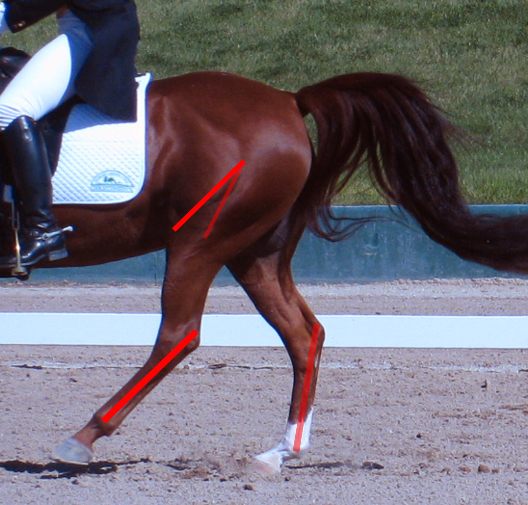


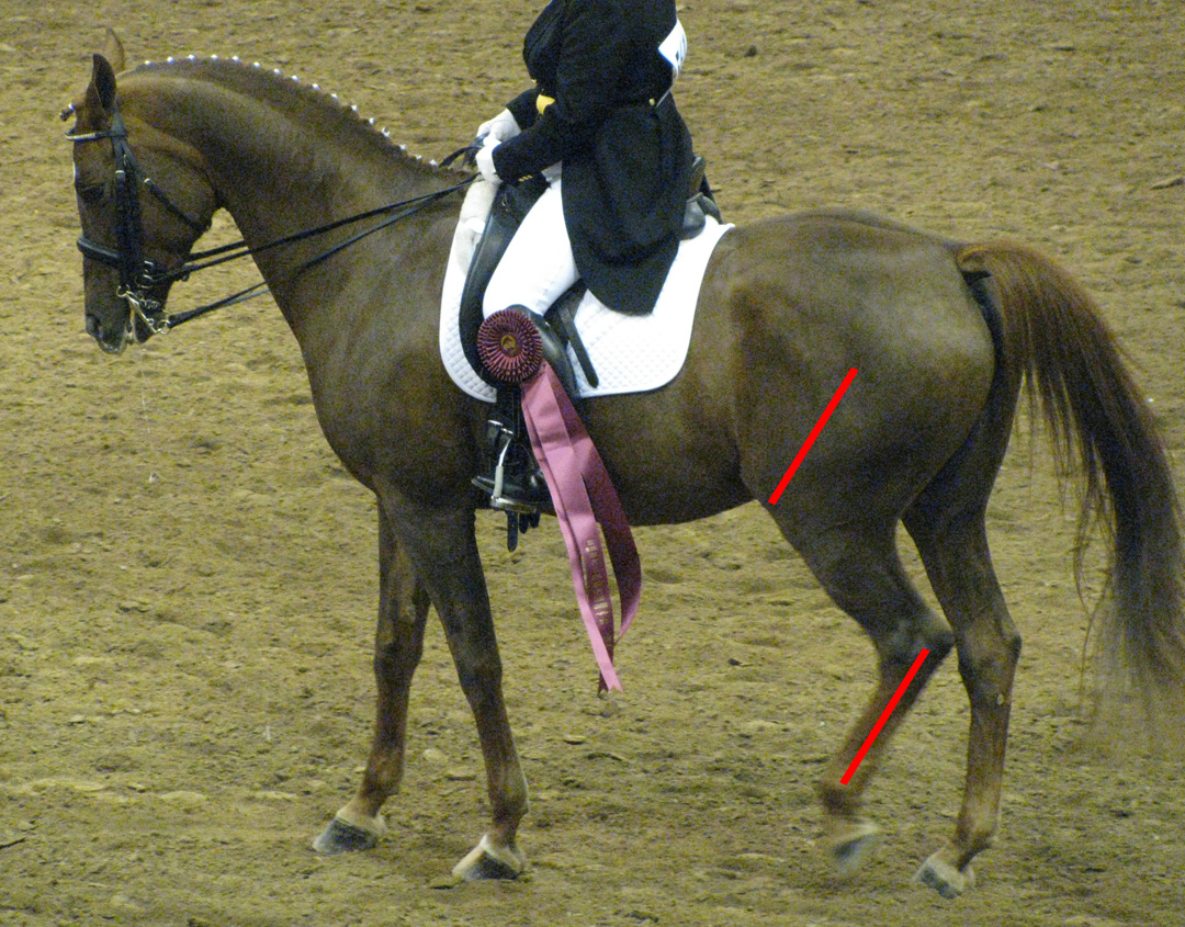

Like the foreleg, the hindlimb even more so moves like a drafting lamp. Note the orientations of the femurs and metatarsals along with the scapulae and the radii, and the humeri and the metacarpals.
Another common error is a straight plane during hindlimb flexion that misinterprets the anatomical structure and biomechanical function of the stifle and hock. Seen from the front, the equine hindlimb cannot flex on a straight plane due to the "popping out" of the stifle and the spiral construction of the hock. Only in extension does the hindlimb straighten out, more or less. Likewise, when the sculpture depicts a standing horse, the hindlimb is usually facing forward on a straight plane (as so often erroneously illustrated in conformation texts) when, in fact, it should be slightly oriented outward as a whole unit, from stifle to toe, away from the median. This is the correct, natural angulation of the hind leg and shouldn't be confused with sickle-hocks. When the hind leg is oriented straight forwards from stifle to toe, the hind leg is actually bow-legged, a conformational fault.
Other common faults are found in the misinterpretation of the stifle, hock, fetlock and foot joints in function or structure, creating mistaken biomechanics, misshapen forms, or incorrect topography. A lack of symmetry from the hock down can also be found, most notably in the fetlock joint and the structure and articulation of the foot structures and joints. The hock is also typically flawed in topography, having "lumps and bumps" in the wrong locations, either seen from the side, or from the front and back. More often, the hock is indistinct and puffy, lacking the necessary lumps n' bumps altogether. Additionally, the hocks's calcaneum is also often too pointy or not moving in synch with the metatarsal, being curved either upwards or downwards away from its proper alignment. Sometimes a flexed hock will be compressed in the front aspect, as through the bones themselves were squished. However, the hock bends by leveraging, not by compression.


Seen from the back, the calcaneum and associated tendons and ligaments down the back of the leg are also often mistakenly the same width instead of the subtle curve and width of the calcaneum in relation to its surrounding structures. In other words, there's no "bulb" to indicate the tip of the calcaneum. Hindlimb's can also be pathological, having puffiness, swellings, depressions, or asymmetries where none should be found. Also found is a misunderstanding of the structure of the tibia, cannon, and pastern bones as seen in a curvature of these bones, often referred to as "spaghetti legs," which we see in the forelimb as well, at times.


These photos demonstrate the natural and normal outward angulation of the hindlimb. The hindlimb should never be oriented on a straight forwards plane when standing.
Seen from the back, the calcaneum and associated tendons and ligaments down the back of the leg are also often mistakenly the same width instead of the subtle curve and width of the calcaneum in relation to its surrounding structures. In other words, there's no "bulb" to indicate the tip of the calcaneum. Hindlimb's can also be pathological, having puffiness, swellings, depressions, or asymmetries where none should be found. Also found is a misunderstanding of the structure of the tibia, cannon, and pastern bones as seen in a curvature of these bones, often referred to as "spaghetti legs," which we see in the forelimb as well, at times.

The hindleg is set towards the outside of the hindquarter, not in the middle.
Sometimes we also see hind legs oriented in the middle of it's haunch, as though the bulk of the Gracilis didn't exist. This causes a narrow hind end and stance. Instead, the hind legs are oriented towards to outside of the haunch, away form the median.
Furthermore, a lack of adequate physics in motion is typical, with the hindlimb not sufficiently demonstrating the power of thrust, weight-bearing, or stopping necessary to best mimic the effort we see in life. We can sometimes see this in the fetlock joint not being depressed downward, but instead the foot bones are "perched" like a bird on a branch in relation to the ground.
Furthermore, a lack of adequate physics in motion is typical, with the hindlimb not sufficiently demonstrating the power of thrust, weight-bearing, or stopping necessary to best mimic the effort we see in life. We can sometimes see this in the fetlock joint not being depressed downward, but instead the foot bones are "perched" like a bird on a branch in relation to the ground.
We also find a confusion in the topographical planes of the hindlimb, most notably those planes falling away from the femur and stifle as well as those of the gaskin and especially the hock. Musculature can be confused, especially in how the hindquarter musculature merges with that of the gaskin. Gaskin muscles are also often too bulbous or engaged when they should be relaxed, which can be seen in some extended hindlimbs. Muscles can be carved in for delineation rather than indicated by soft curves and the 3D quality of muscle masses. In other words, the muscles of the hindquarter are defined by lines, or gouged tracing rather than fleshy indications; the musculature expresses more as soft troughs and bulges rather than harsh lines. Think of bulk rather than defined lines indicating muscle. The muscles of the hindquarter also undergo a great deal of morphing in motion, so we shouldn't see "standing" musculature on a piece depiction motion, and visa versa. Likewise, standing muscle definition and delineation is too often seen on hindlimbs that depict motion. Remember, the horse's muscles don't manifest like an articulated anatomy chart...they morph and contort, merge, goo, and pooch in response to articulation and physics.


Biological Aspects to Consider about the Hindlimb
The equine hindlimb was designed by nature for thrust and propulsion while the forelimbs are designed more as rods, pole-vaulting the forequarter forwards in motion, especially in the gallop. This gives the animal a springy, powerful stride and contributes to that look of hovering flight. And the faster the animal goes, the more important that coordination of the pole-vault motion of the forelimbs becomes. This is why horses trip in the forelimbs whereas they fall out from under themselves in the hindlimbs. It's also why horses forced to move too slowly or out of synch with the coordination of a normal gait can plod, trip, or kick up dirt with their fore hooves because the pole-vaulting effect of the forelimbs cannot be adequately exploited by the hindlimbs. We can sometimes see this with "peanut-rollers" or Western Pleasure horses made to move with impure gaits.
Furthermore, motion that's consistent to natural equine coordination and movement will preserve the integrity of the hind leg joints, even under extreme performance demands. Injuries and pathologies of the hindlimb occur when the animal is forced into unnatural postures that alter natural coordination and function, which can lead to puffiness, bumps, and swellings. We often see this in dressage as so many horses are forced into false collection due to rollkur and "push-pull" or "frame" riding. Indeed, false collection can lead to many pathologies in the hindlimb from String Halt to being "strung out" behind, to even ruptured stifle ligaments.
Furthermore, motion that's consistent to natural equine coordination and movement will preserve the integrity of the hind leg joints, even under extreme performance demands. Injuries and pathologies of the hindlimb occur when the animal is forced into unnatural postures that alter natural coordination and function, which can lead to puffiness, bumps, and swellings. We often see this in dressage as so many horses are forced into false collection due to rollkur and "push-pull" or "frame" riding. Indeed, false collection can lead to many pathologies in the hindlimb from String Halt to being "strung out" behind, to even ruptured stifle ligaments.
The hindlimb also moves fast. Those legs positions and push off occur in the blink of an eye and the forelimbs "catch" the forward motion to support the forequarter. Capturing this kind of nimble movement is important for an equine sculpture since a "clunky" feel can really "stop" the motion visually.
Conclusion to Part XIII
Phew! That was a lot to chew on, wasn't it? The hindlimb maybe be a bit easier than the forelimb, but it's complicated nonetheless. So many details to factor into our equation! But how we sculpt the hindlimb can mean the difference between a convincing sculpture and one that's not since the physics of impulsion influence them so greatly. If done right, they can really capture the idea of mass, thrust, and force, as well as fleeting agility so characteristic of this animal's motion.
So now that we have this last aspect under our belt, let's delve into details in the next installment. The equine has lots of them that deserve our careful attention, plus they're fun to infuse into our work because they really bring a piece alive.
So until next time...propel forwards in your understanding equine biology!
"The better an artist can mimic how the eye sees, the more effective and natural the paintings will be."
So now that we have this last aspect under our belt, let's delve into details in the next installment. The equine has lots of them that deserve our careful attention, plus they're fun to infuse into our work because they really bring a piece alive.
So until next time...propel forwards in your understanding equine biology!
"The better an artist can mimic how the eye sees, the more effective and natural the paintings will be."
~ Kenn Backhaus


